
6-12 Months, Augma Bone Cement Academy, Bone Cement Expert, Clinical Cases, Clinical Indication, Clinician, Coverage Used, Dental Notation, Images, Lower Left Molar, Media, Peri-Implantitis, Post-Op Period, Soft Tissue Augmentation, Video, Wound Dressing
Rehabilitation of Large Bone Defect in the Left Posterior Part of the Mandible
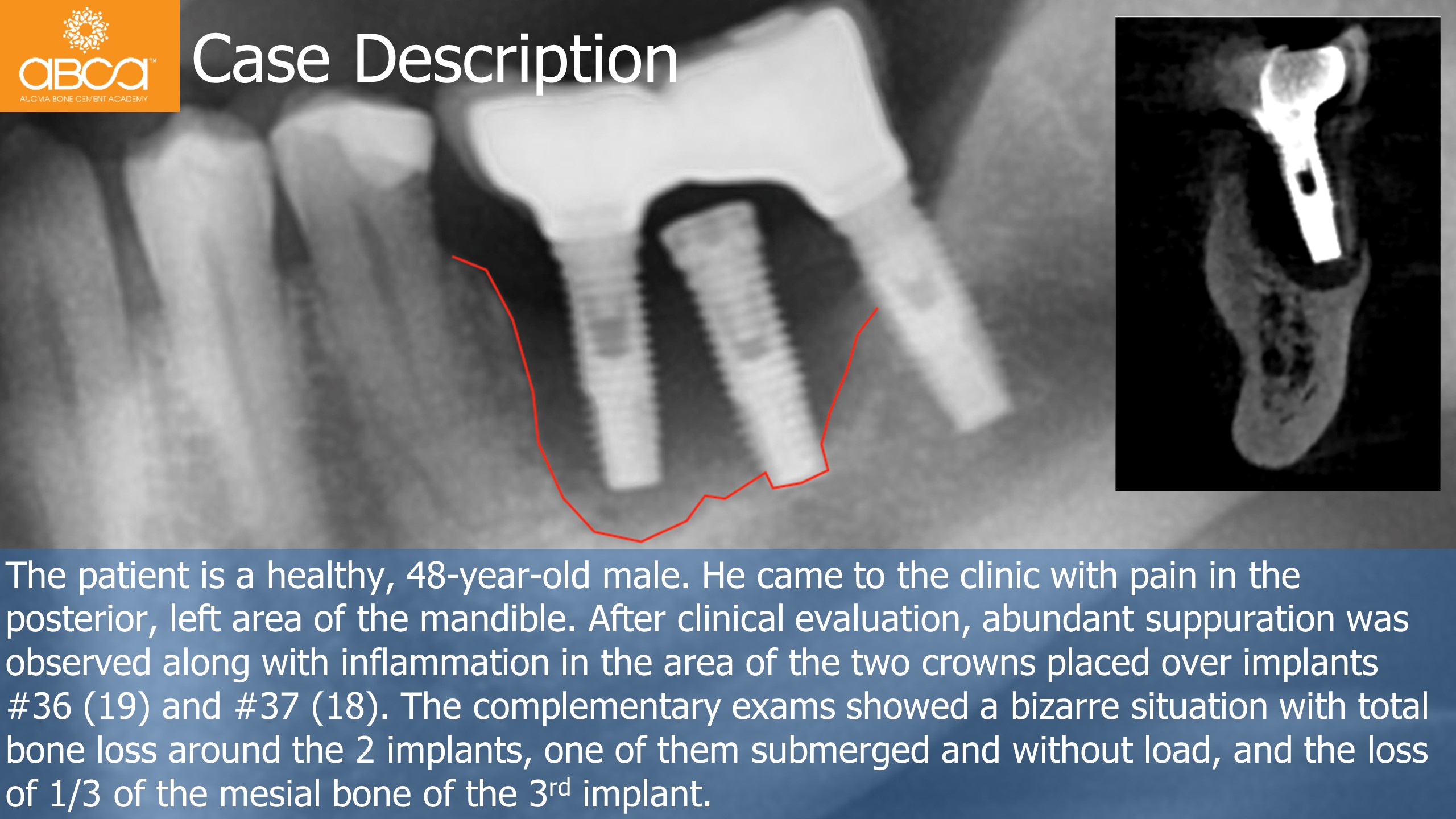
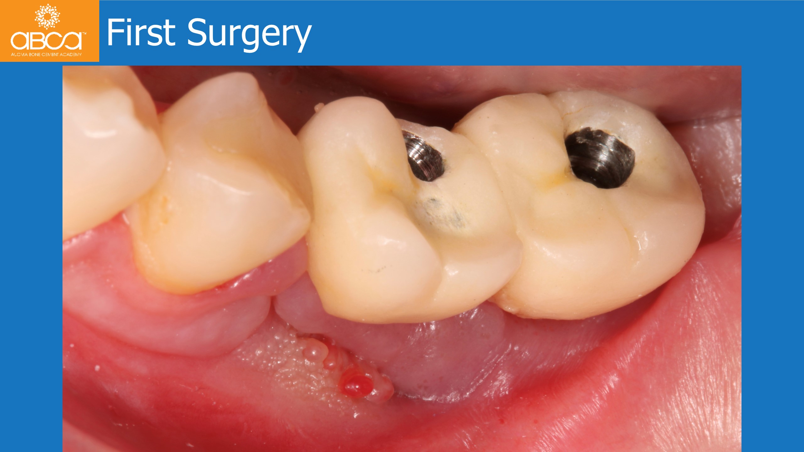
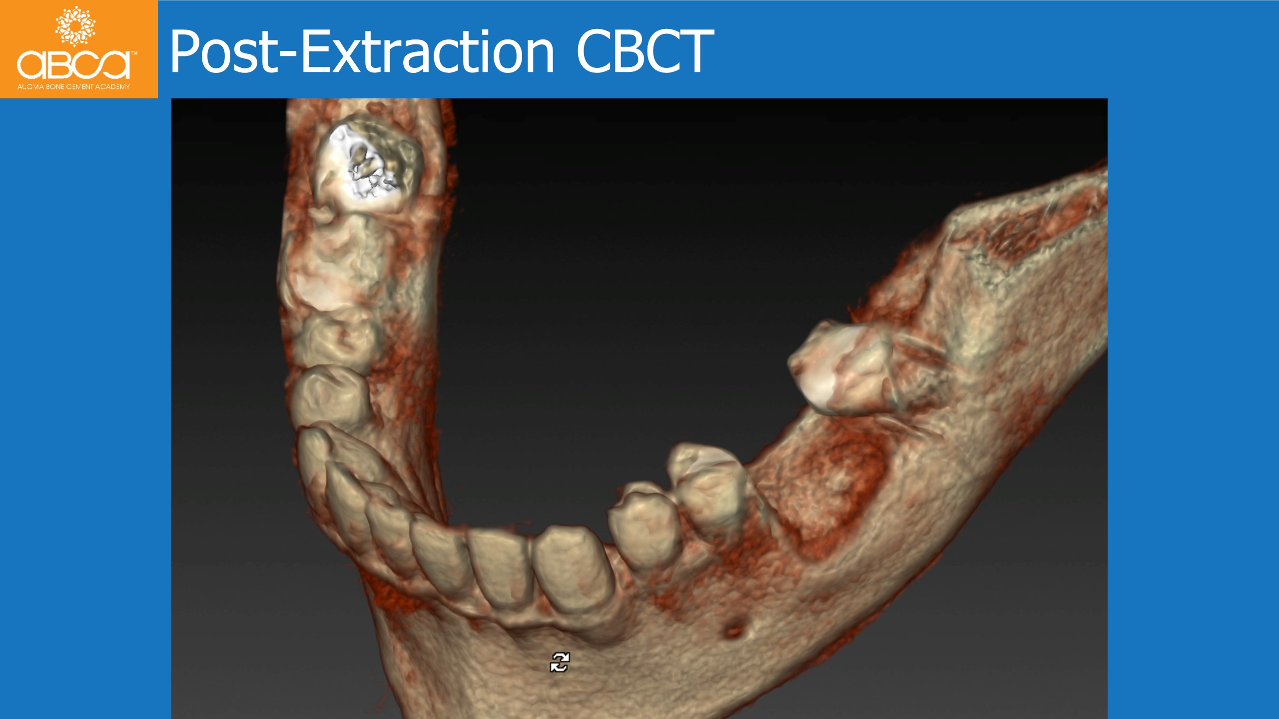
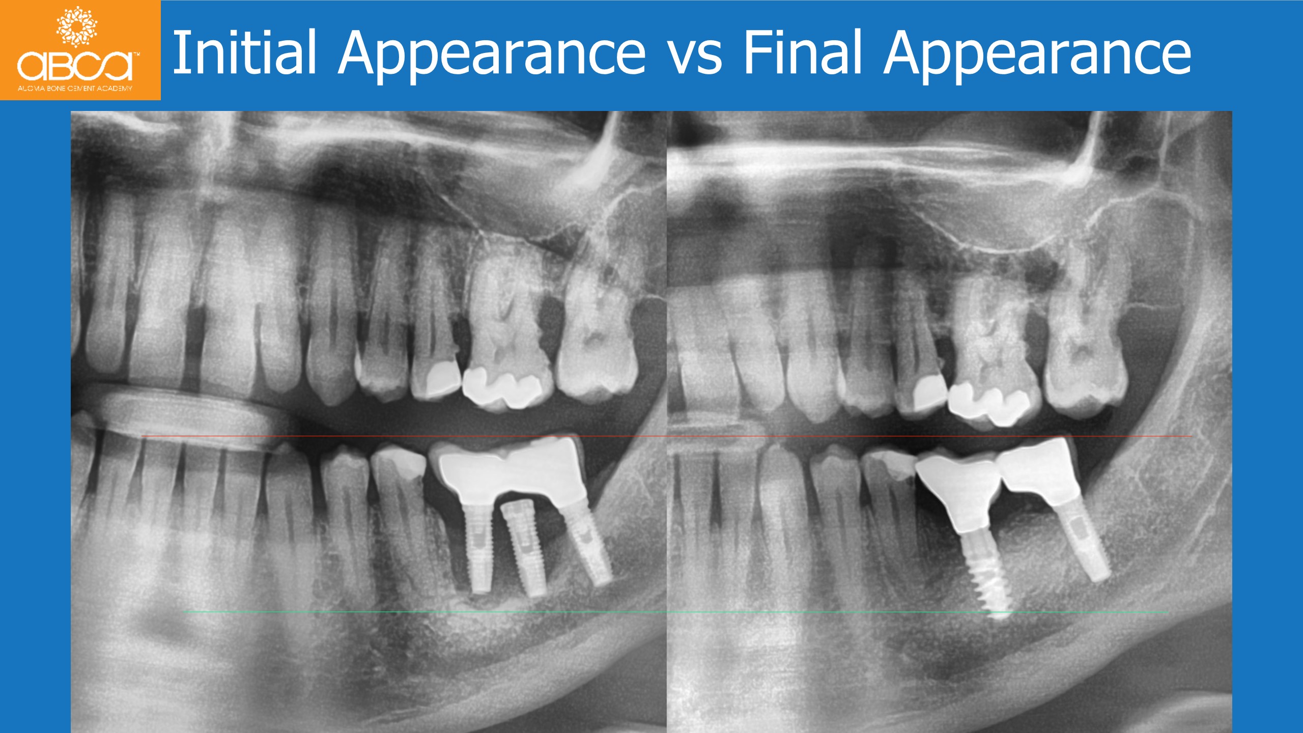
The patient is a healthy, 48-year-old male. He came to the clinic with pain in the posterior, left area of the mandible. After clinical evaluation, abundant suppuration was observed along with inflammation in the area of the two crowns placed over implants #36 (19) and #37 (18). The complementary exams showed a bizarre situation with total bone loss around the 2 implants, one of them submerged and without load, and the loss of 1/3 of the mesial bone of the 3rd implant.
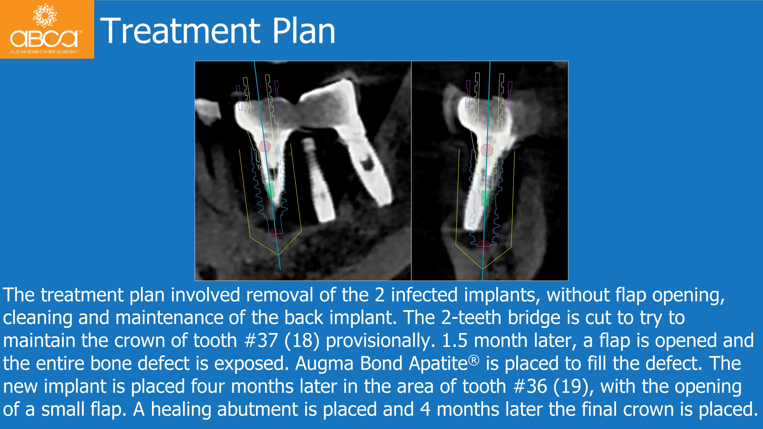
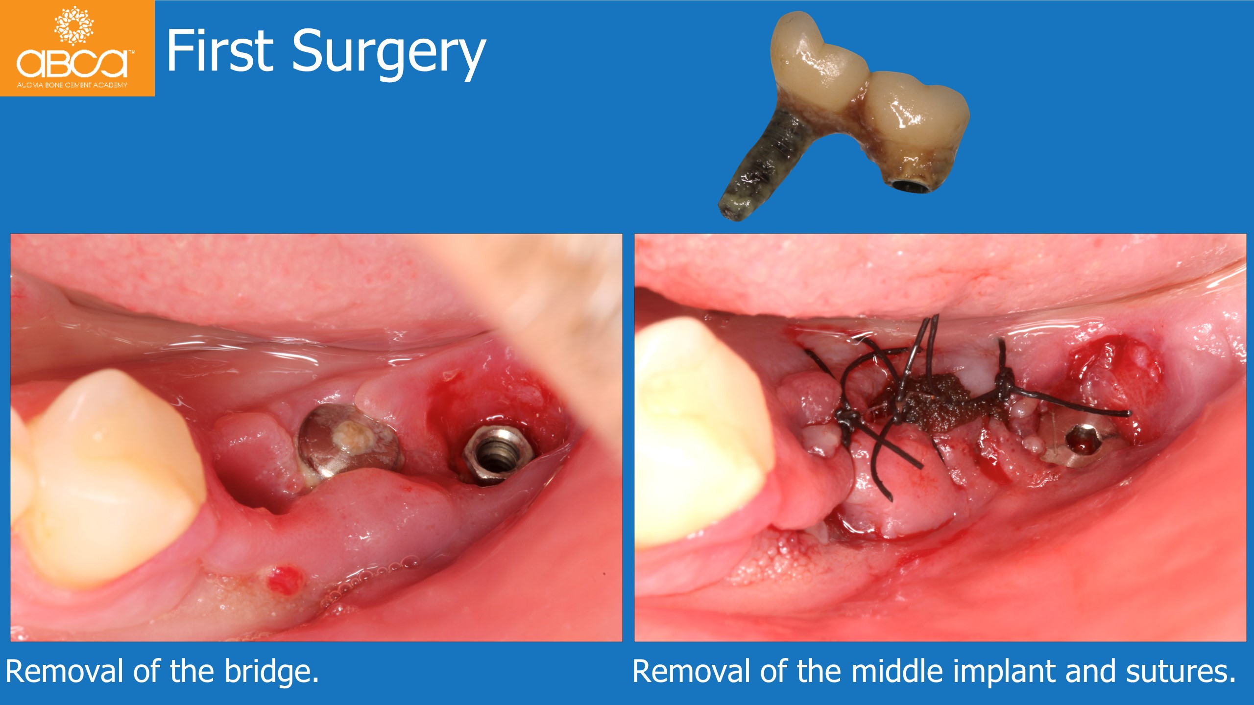
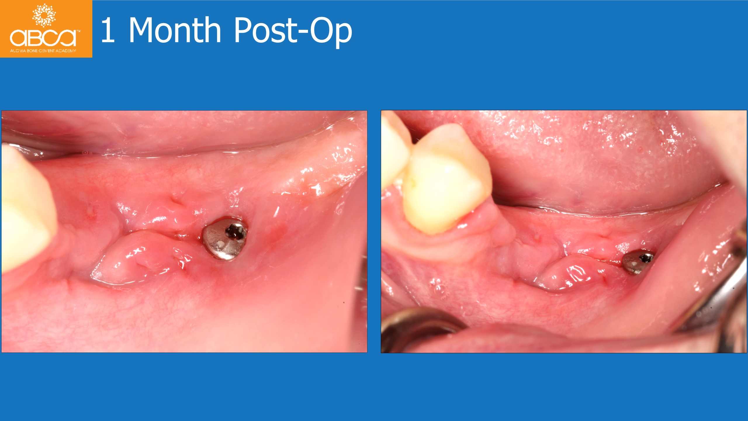
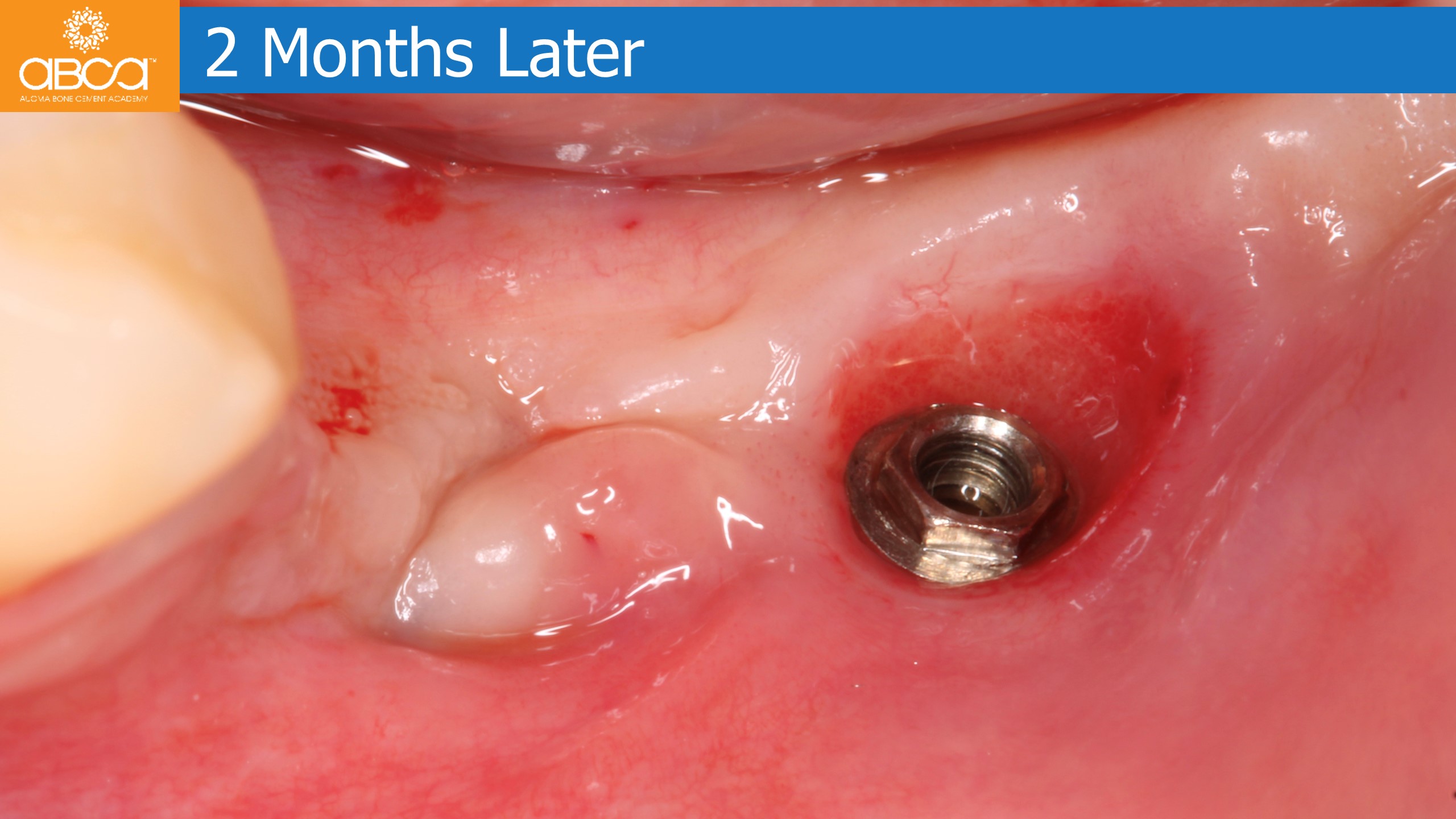
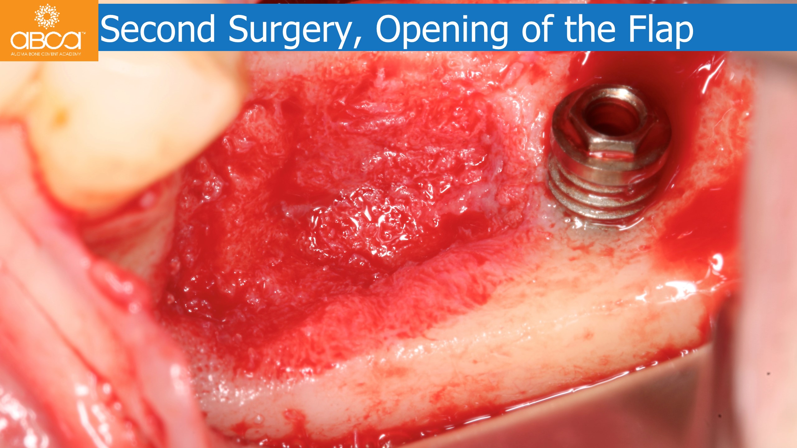
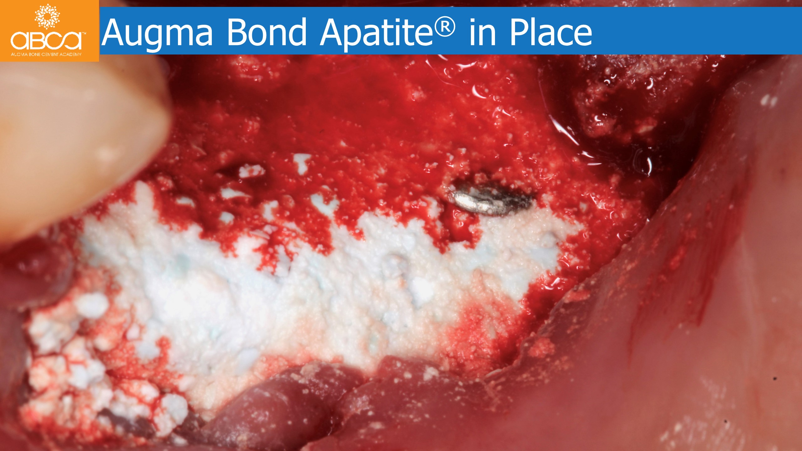
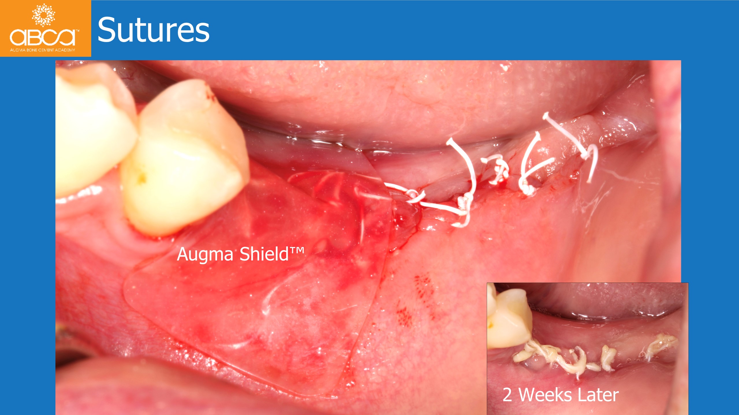
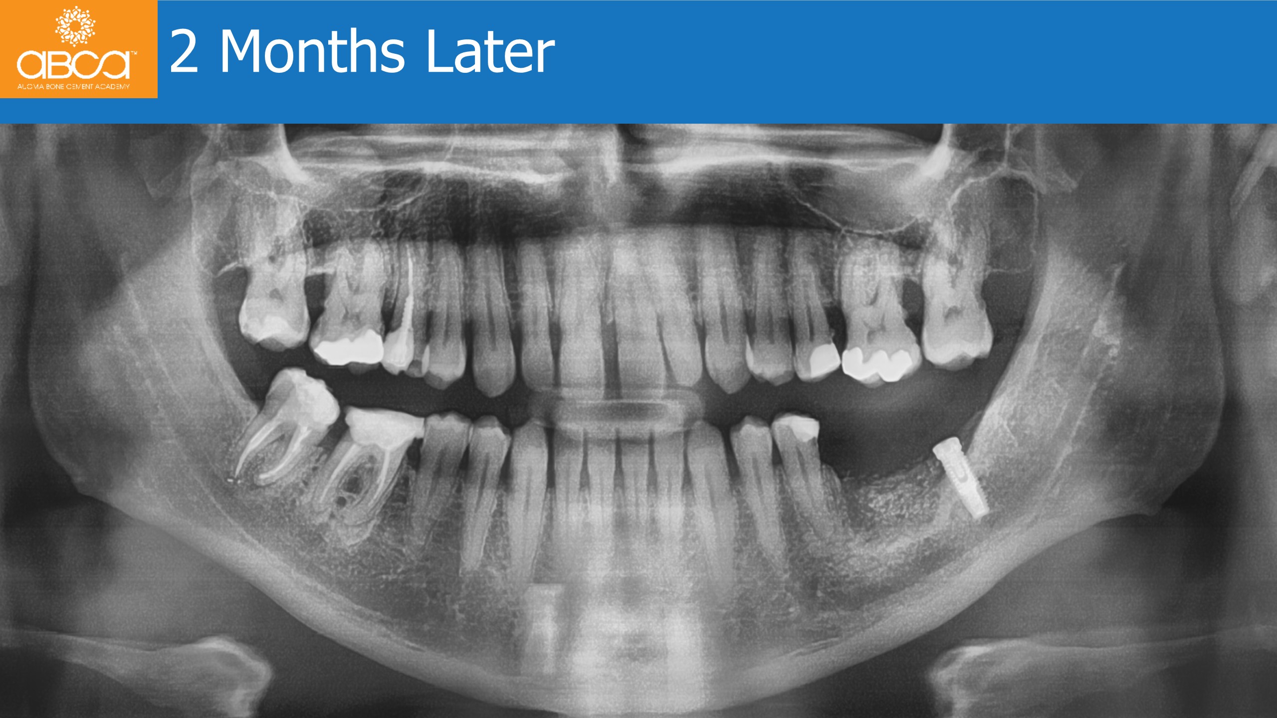
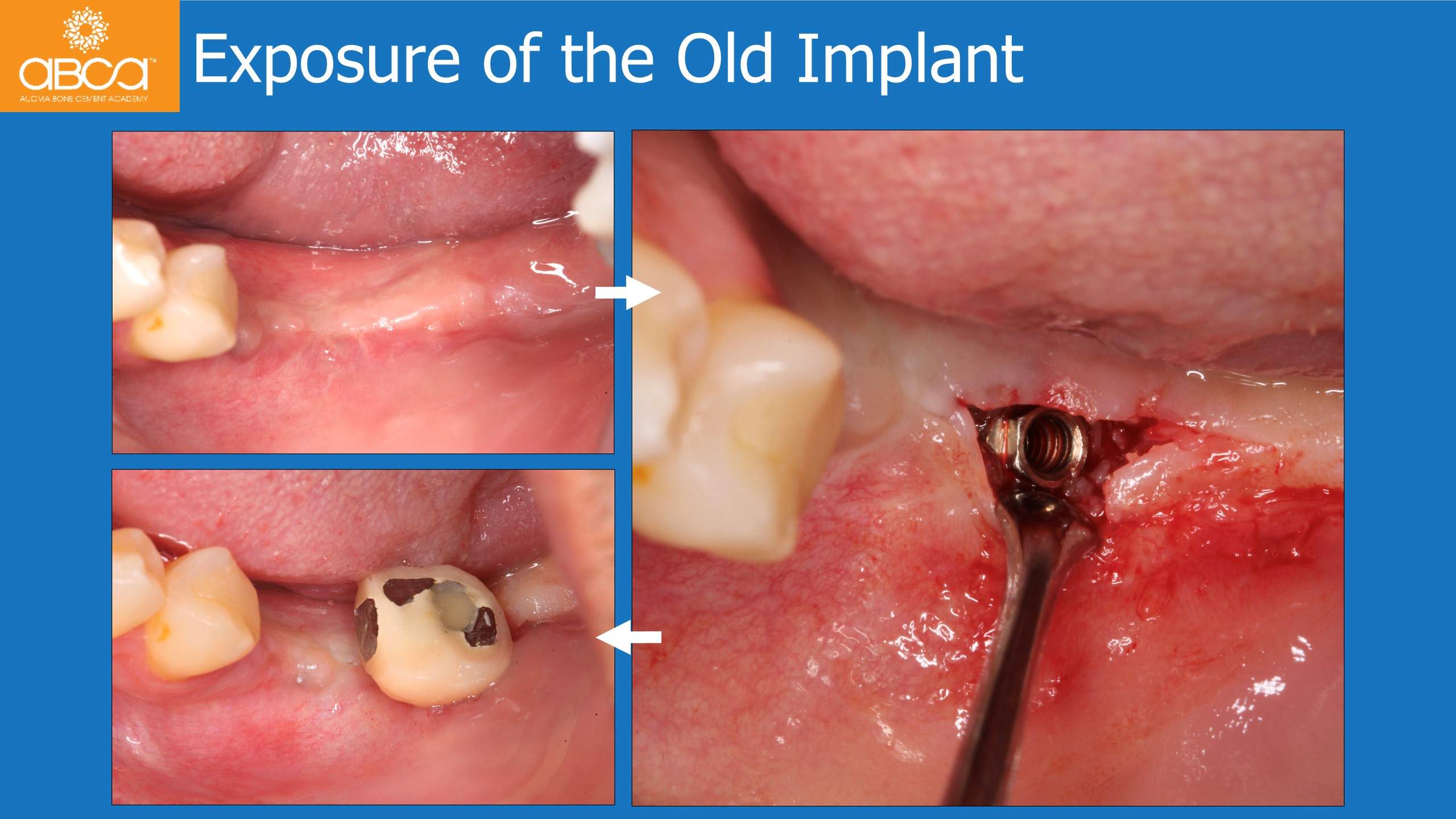
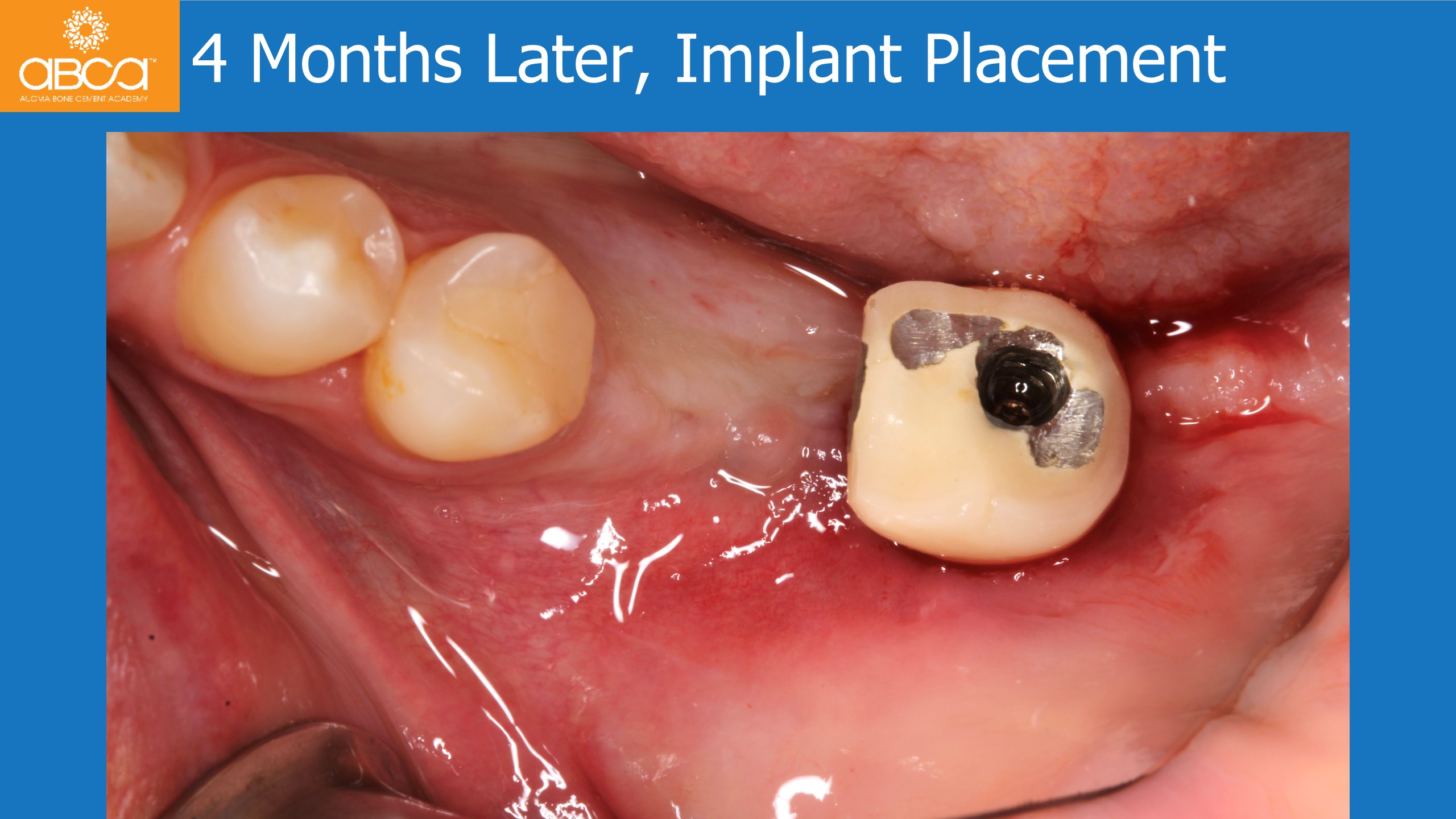
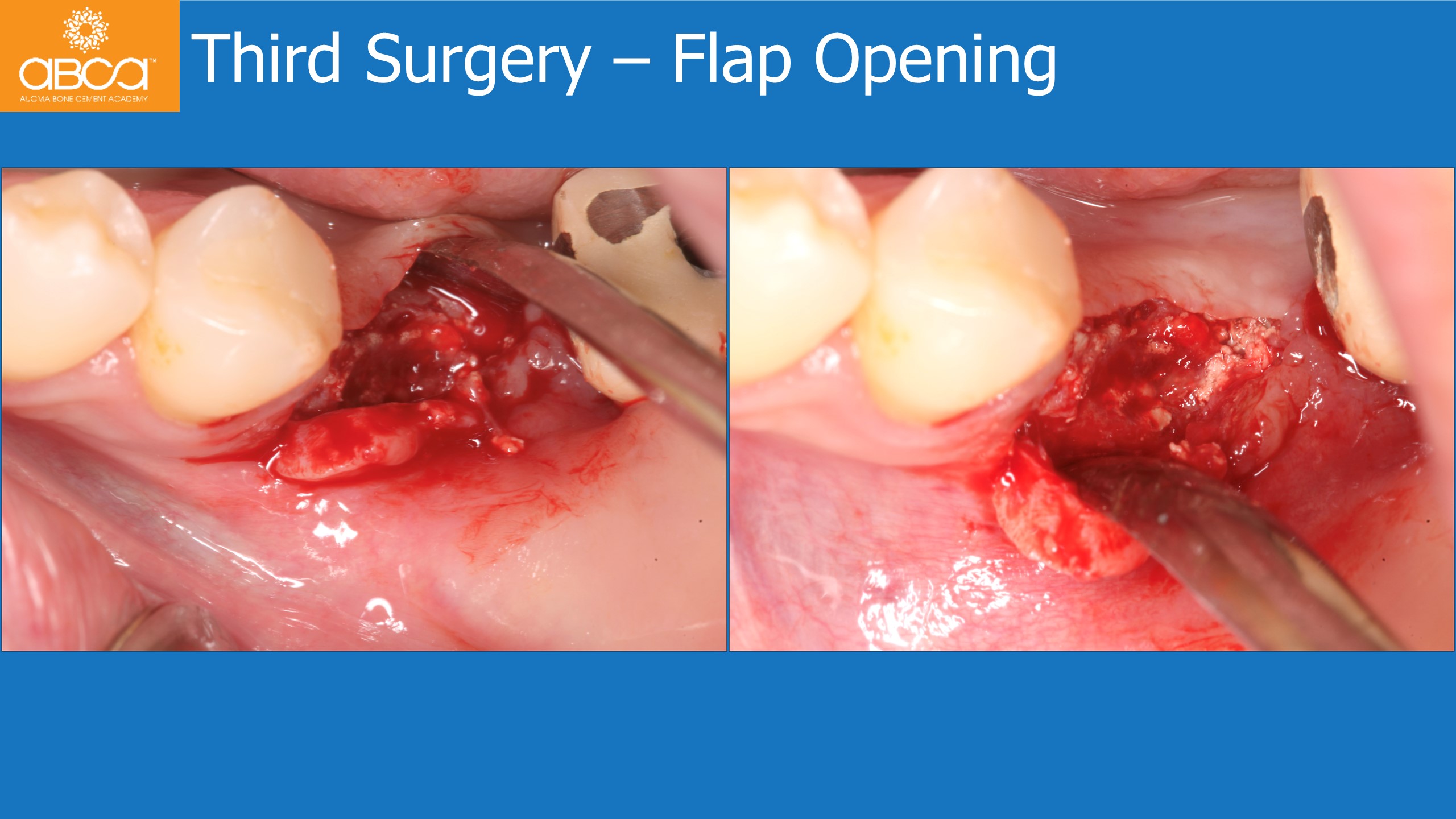
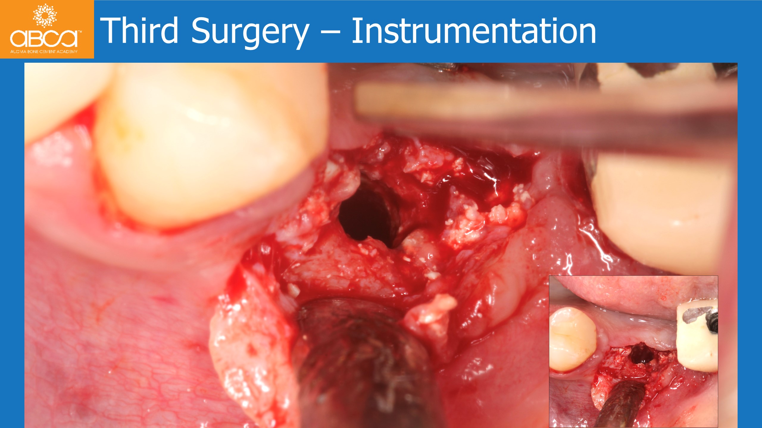
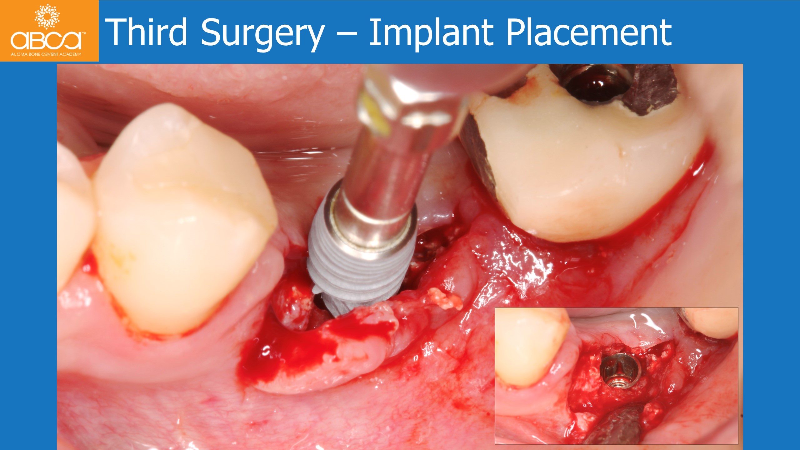
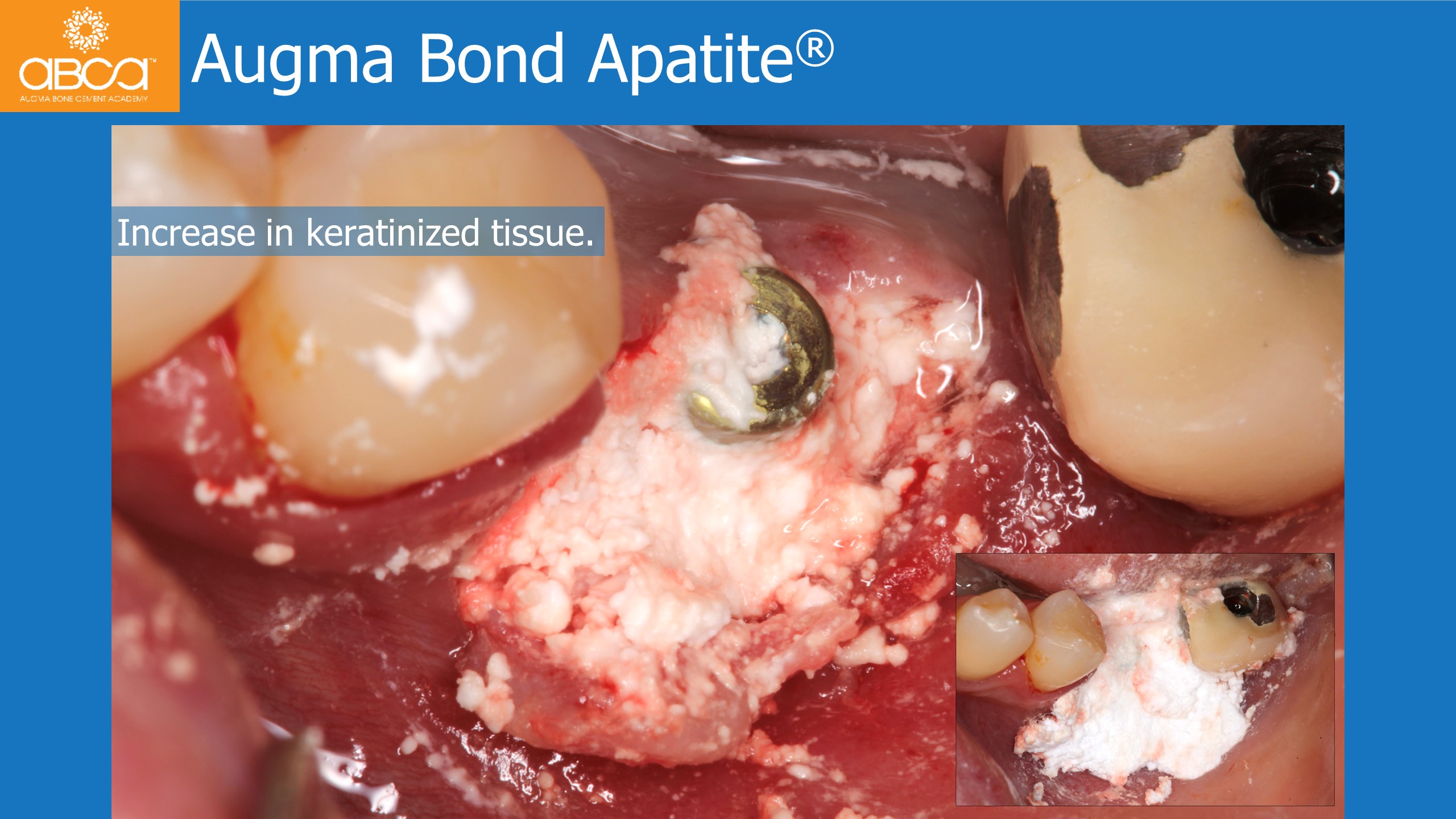
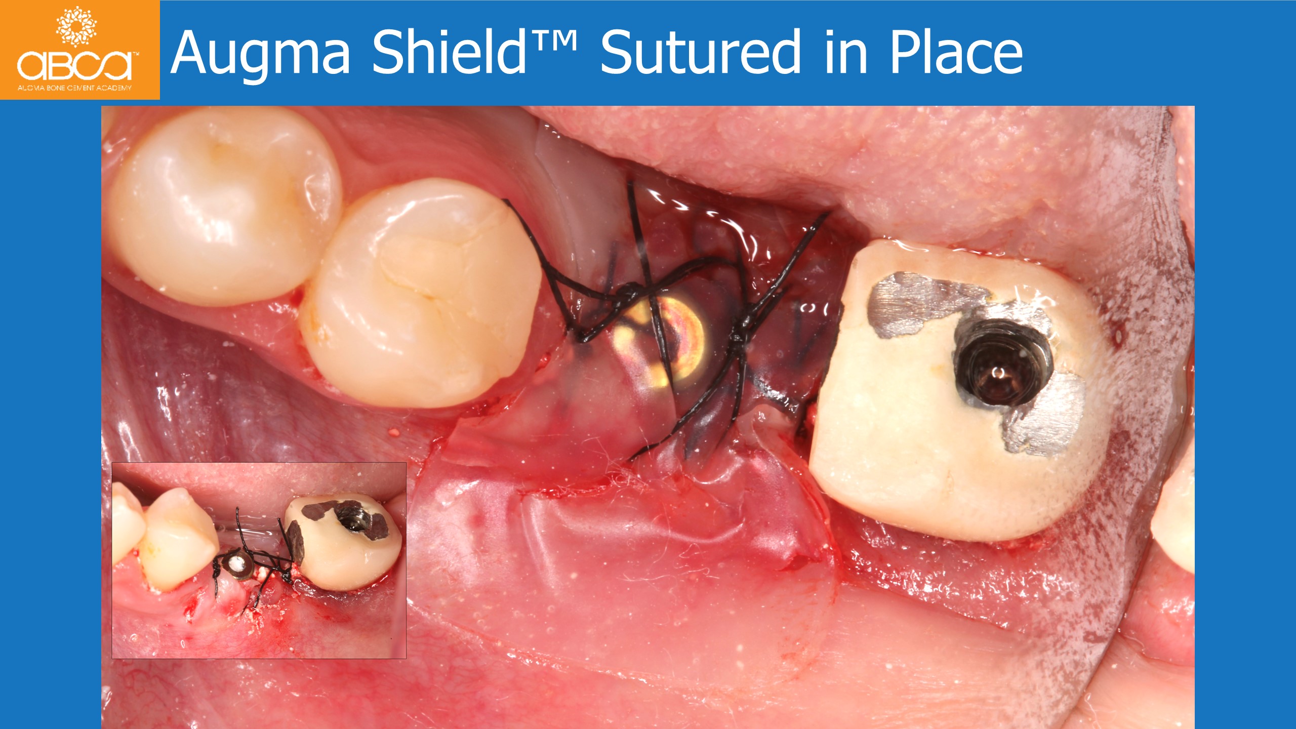
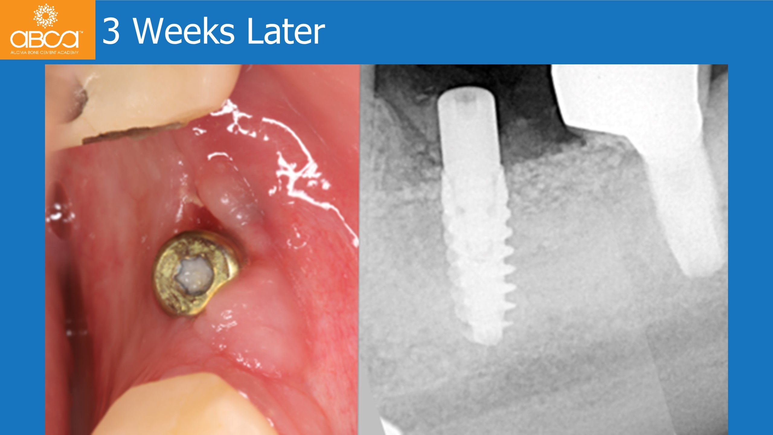
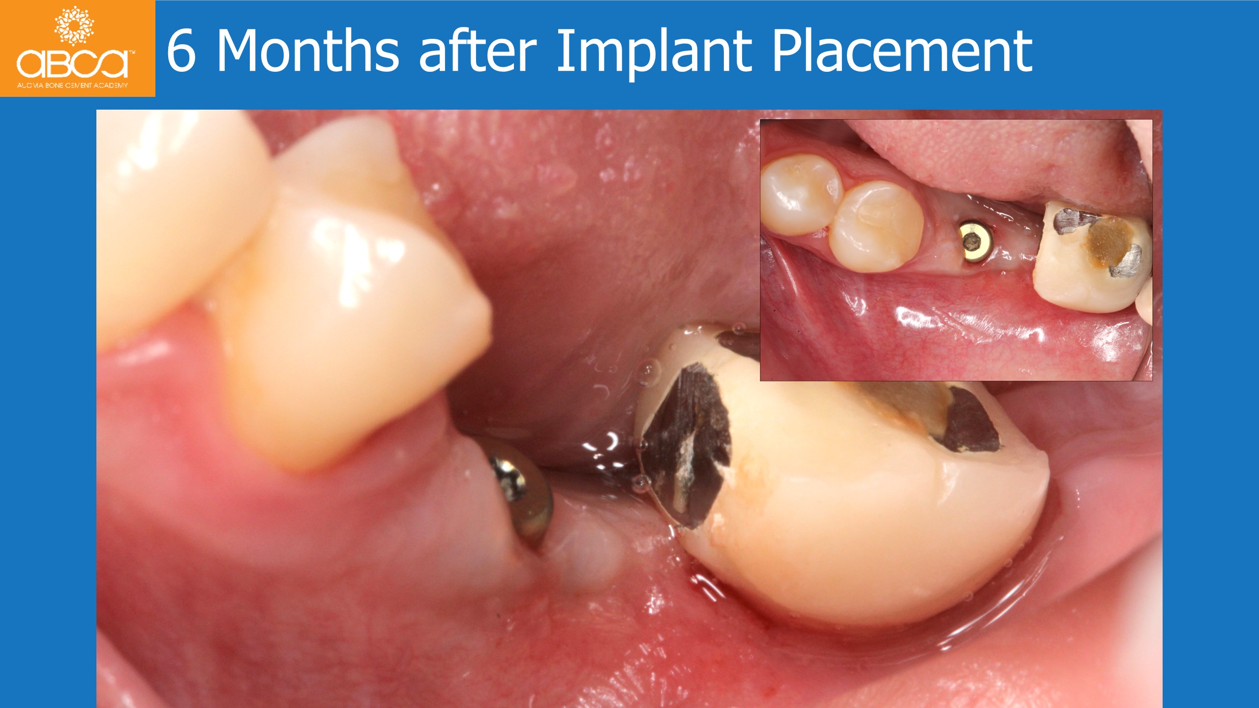
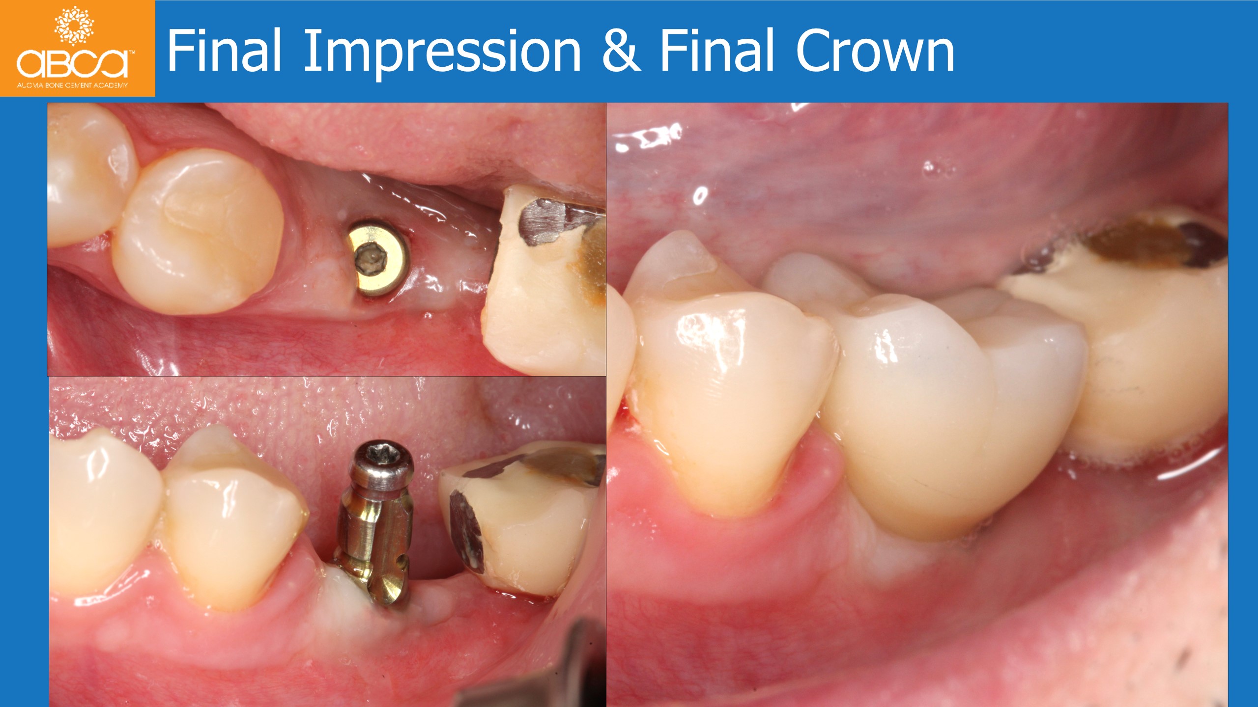
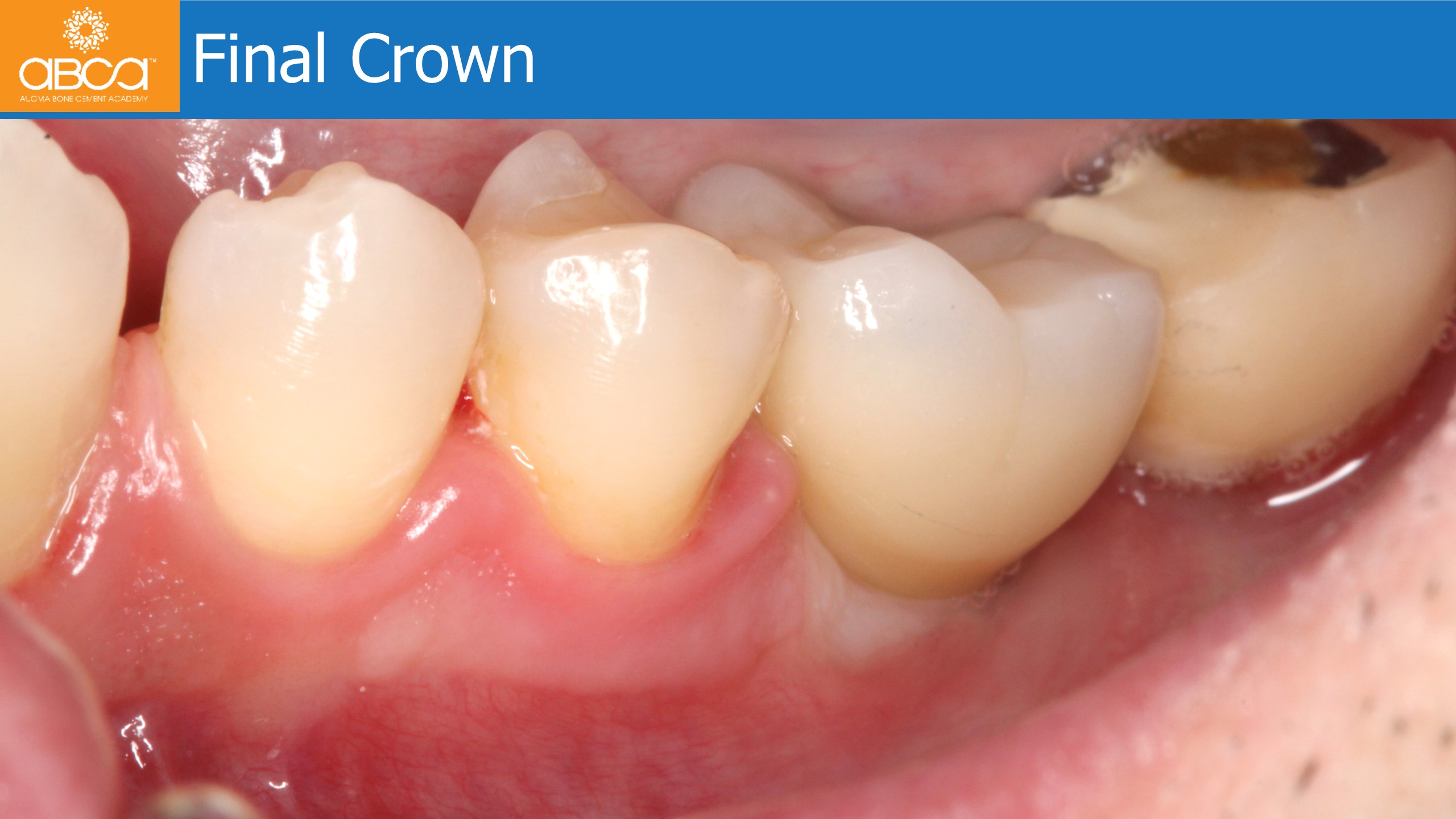
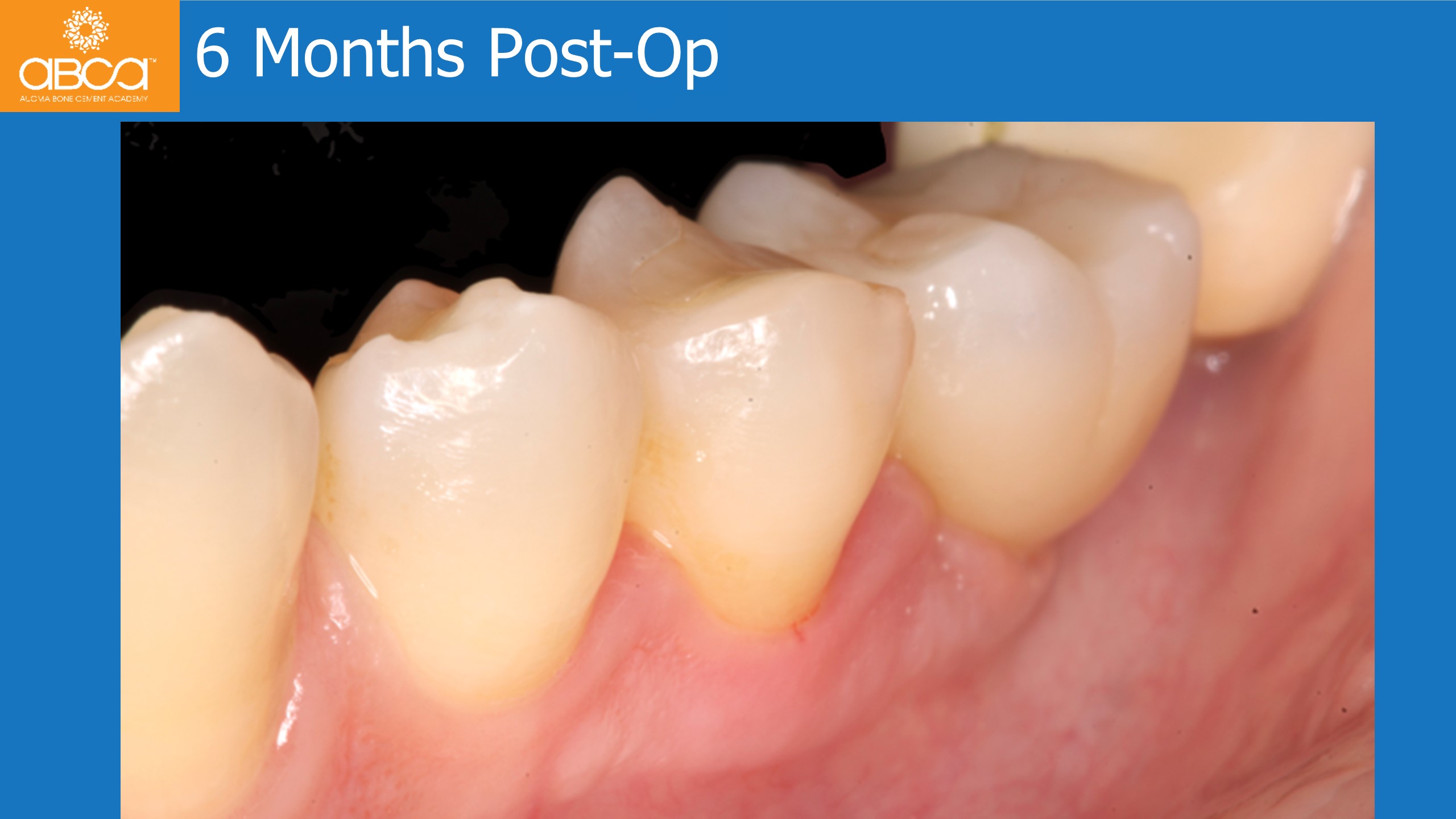
The treatment plan involved removal of the 2 infected implants, without flap opening, cleaning and maintenance of the back implant. The 2-teeth bridge is cut to try to maintain the crown of tooth #37 (18) provisionally. 1.5 month later, a flap is opened and the entire bone defect is exposed. Augma Bond Apatite® is placed to fill the defect. The new implant is placed four months later in the area of tooth #36 (19), with the opening of a small flap. A healing abutment is placed and 4 months later the final crown is placed.